VisioVIEWPowerfulImageViewingandAnalysisTools
Unlock Precision in Veterinary Imaging
The VisioVIEW DICOM Viewer bundle is crafted with veterinary professionals in mind, providing an easy-to-use platform for managing, viewing, analyzing, and interpreting medical images from CT, MRI, X-ray, ultrasound, and more.
Key Components of VisioView
Key Components of VisioView
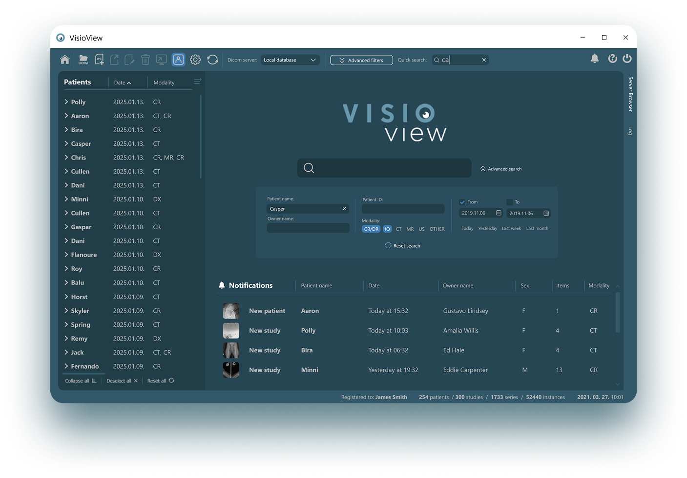
Study Browser
The DICOM files present on the PACS are represented in VisioVIEW via a Study Browser. This highly optimized interface offers great scalability based on the size of the clinic.
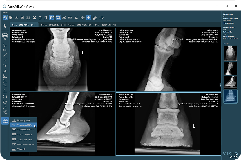
Advanced DICOM Viewer
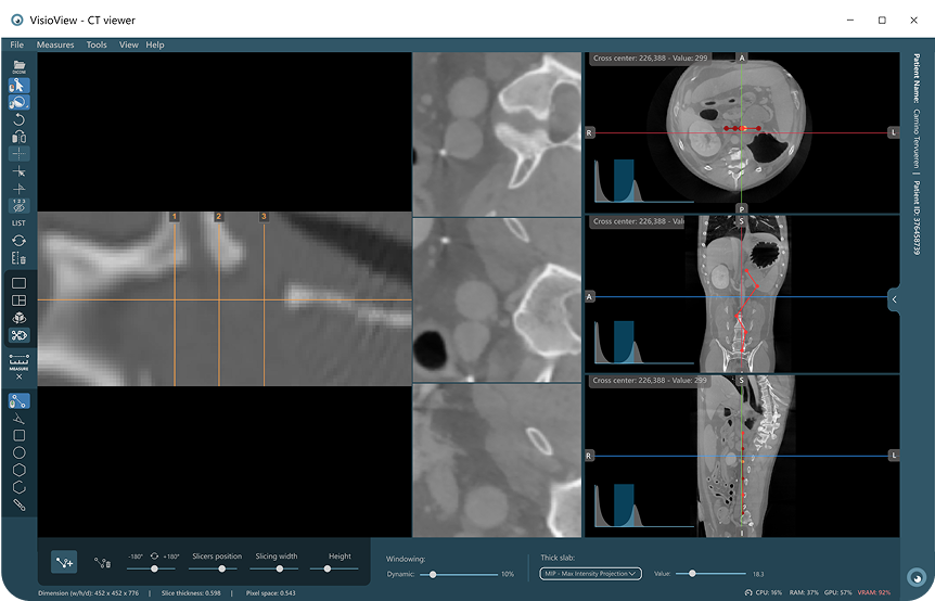
CT Viewer with MPR and 3D
DICOM Image Viewing and Manipulation
Designed specifically for veterinary care, VisioVIEW delivers quick access to detailed image analysis.
Multi-Modality Support
Seamlessly handles multiple imaging modalities, including MRI, CT, X-ray, ultrasound, and more.
Windowing & Leveling
Adjust contrast and brightness to highlight specific areas of interest, enhancing the visibility of details in complex images.
Cine Mode
View image sequences or series (CT or MRI scans) in a video-like format, simplifying the analysis of dynamic data.
Advanced 3D Imaging Capabilities
Transforming CT and MR Studies into Detailed 3D Insights
Multiplanar Reconstruction (MPR & CMPR)
Enable 3D views from 2D slices, with axial, sagittal, coronal and custom curved reconstructions for enhanced spatial understanding.
3D Volume Rendering
Provides volumetric visualization of structures, offering advanced 3D perspectives for precise analysis.
Precise Tissue Differentiation
The HU Distribution Editor enables precise tissue visualization by customizing opacity, color, and HU ranges for specific tissues.

Advanced Annotation & Measurement Tools
Enhance Precision with Comprehensive Analysis Tools
Basic Measurements
Offers a suite of tools for distance, angle, area, and circumference measurements, essential for accurate analysis.
Annotations
Add markers, text, and highlights directly on images to specify important findings or observations.
Region of Interest (ROI)
Define and measure specific areas within the images to focus on critical regions for more precise assessment.
Zoom and Rotate
Basic manipulation tools for adjusting images to fit analysis needs.
Hounsfield Unit
Especially useful in CT scans to access the density of tissues.
Image Comparison
Side-by-side comparison of multiple images, facilitating comparison of studies.
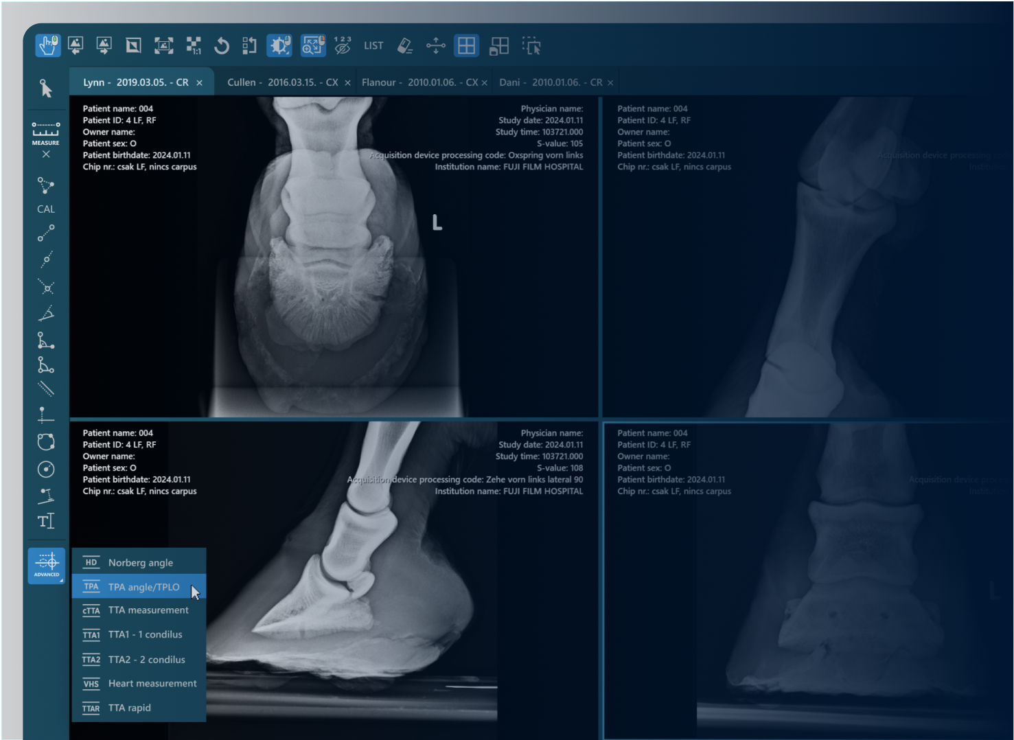
Advanced Multistep measurements
Full support for multi-step advanced measurements, guiding the user through the measurement construction.
Tibial Plateau Angle
Tibial Plateau Leveling Osteotomy
Tibial Tuberosity Advancement
Tibial Tuberosity Advancement
Tibial Tuberosity Advancement
Norberg Angle
Vertebral Heart Score
Cobb Angle
Hip Distraction Index

Multi-User Collaboration & Teleradiology Support
Seamless Collaboration and Remote Accessibility
Concurrent Access
Enable multiple users to access and edit images or studies simultaneously, enhancing collaboration between veterinary professionals.
Remote Access
Access PACS servers over the internet, enabling veterinary professionals to review studies from anywhere, at any time.
Email Support
Share and communicate image data directly via email, with configurable settings for easy integration with services like Gmail.
Image Gallery Presentation
Create and send curated image galleries directly from VisioVIEW, perfect for patient consultations or sharing study results.
DICOM Hanging Protocols
Customize DICOM Hanging Protocols to meet specific clinical needs
Study and Instance based rules for precise targeting
Adjust view sizes, image sequence order, and more
Assign specific layouts locally based on viewer state


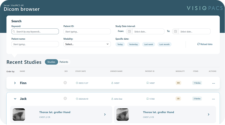
Optimized Study Browser
Customizable Browsing for Efficient Study Access
Scalability
Whether for small or large veterinary clinics, VisioVIEW's Study Browser allows you to scale and organize your imaging data efficiently. Filter and sort by DICOM parameters to find studies quickly.
Advanced filtering
Large clinics can opt to display only a subset of their images, while still having the ability to search for specific studies as needed.
DICOM management
VisioVIEW ‘s Study Browser empowers users with full control over their imaging data. Users can delete or import studies and DICOM files, connect to remote PACS servers, and even edit DICOM metadata.

Integration & Interoperability
Connectivity Across Systems and Platforms

PACS/DICOM Integration
Directly retrieve studies from PACS servers via DICOM Query/Retrieve for seamless access to imaging archives.
DICOM-CD/DVD Support
Load DICOM images from CDs or DVDs provided by patients or other veterinary practices, ensuring accessibility without PACS.
DICOMDIR Support
Read and navigate DICOMDIR files for easier management and retrieval of stored studies from structured directories.
Export to Common Formats
Effortlessly export images in formats like JPEG, BMP, and PNG, ideal for sharing with other professionals or for inclusion in reports.
Export Series as Video
Export dynamic image sequences (such as CT or MRI cine sequences) as video files (MP4) for easy sharing and patient communication
Burn to CD/DVD with Viewer
Offer the option to export studies bundled with the VisioVIEW viewer for offline access, ensuring the recipient has everything needed for proper viewing.
AWS Support
Send DICOM files and exported images as compressed ZIP files via AWS shared download links for efficient, cloud-based file management.
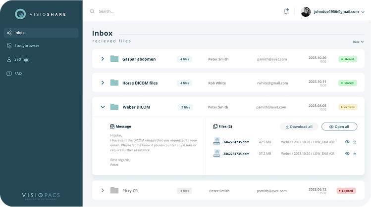
VisioSHARE - Effortless DICOM Sharing
VisioSHARE is a powerful web application tailored for veterinary physicians to securely share DICOM files and medical images with ease.
Make VisioVIEW your VisioVIEW
Personalize your workspace with theme, color and UI scaling options tailored to your environment and preferences.
Switch between Dark, Light, and Normal modes for optimal visibility.
Choose from multiple color styles to match your workflow mood.
Color themes can be combined with dark, light or normal modes


See how it all works together
Experience the full potential of VisioPACS in action!